Shoulder Instability and Recurrent Shoulder Dislocation
Shoulder instability occurs when the ball of the shoulder joint moves excessively within the socket. In some patients, the shoulder may partially slip out of position, known as a subluxation. In others, the shoulder may completely dislocate and require reduction. The shoulder is naturally a very mobile joint. Its stability depends on the shape of the bones, the labrum around the socket, the joint capsule and ligaments, and the muscles surrounding the shoulder. Damage or looseness affecting any of these structures can allow the shoulder to become unstable.
What keeps the shoulder stable?
The shoulder joint consists of the rounded head of the humerus sitting against the relatively shallow socket of the shoulder blade, known as the glenoid. Several structures work together to keep the shoulder centred:
-
The bones provide the underlying shape of the joint.
-
The labrum is a rim of cartilage around the socket that deepens the joint and provides attachment for the shoulder ligaments.
-
The capsule and ligaments help prevent the shoulder from moving beyond its normal limits.
-
The rotator cuff and other shoulder muscles dynamically control the joint during movement.
When these structures are damaged during a dislocation, the shoulder may remain vulnerable to further instability.
Types of shoulder instability
Shoulder instability is generally divided into anterior, posterior and multidirectional instability.
Anterior shoulder instability
Anterior instability is the most common form and accounts for approximately 95% of traumatic shoulder dislocations. The ball moves forwards out of the socket, usually when the arm is raised and rotated backwards.
Patients commonly describe the shoulder “popping out” during a fall, tackle, collision or overhead sporting activity.
A first dislocation can tear the labrum and stretch or damage the shoulder capsule. In some patients, part of the bone may also be damaged. Repeated dislocations can cause progressively greater injury to the labrum, glenoid socket and humeral head.
Posterior shoulder instability
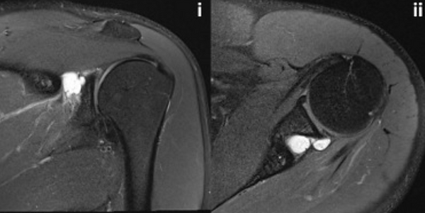
Posterior instability occurs when the ball moves backwards relative to the socket. It is less common and may be more difficult to diagnose. Patients with posterior instability do not always report a complete dislocation. Pain is often the main symptom, particularly during pushing activities such as bench press, push-ups or contact with the arm positioned in front of the body. Posterior instability may be associated with a posterior labral tear. Occasionally, a fluid-filled cyst can form near the back of the shoulder and affect a nearby nerve.
Multidirectional instability
Multidirectional instability describes excessive movement of the shoulder in more than one direction. It is often atraumatic and may affect both shoulders. It is more common in younger patients with generalised joint flexibility or hypermobility. The joint capsule may be unusually loose or capacious rather than having a single traumatic tear.
Physiotherapy is the main treatment for multidirectional instability. Surgery is generally reserved for patients with persistent symptoms despite a prolonged and well-structured rehabilitation program.
What happens when the shoulder dislocates?
A traumatic shoulder dislocation commonly separates the labrum from the front of the glenoid socket. This is often called a Bankart lesion. The capsule and ligaments may also become stretched or torn.
The humeral head can strike the edge of the socket and develop an indentation known as a Hill-Sachs lesion.
Some patients also lose bone from the front of the glenoid. Bone loss becomes increasingly important in patients who have experienced repeated dislocations, long-standing instability or instability during collision sport. The amount and location of bone loss can determine whether a soft-tissue repair is likely to succeed or whether a bone-transfer procedure is required.
Who is at risk of another dislocation?
Age is one of the strongest predictors of recurrence after a first traumatic shoulder dislocation.
Teenagers and young adults have a particularly high risk. Recurrence is reported in approximately 90–95% of patients younger than 19 years and may be almost universal in some patients younger than 16 years.
The risk is also higher in:
-
Contact and collision-sport athletes;
-
Overhead athletes and workers;
-
Patients with glenoid or humeral bone loss;
-
Patients who have experienced several instability episodes;
-
Patients with a large labral or capsular injury; and
-
Patients who wish to return to high-risk sport or work.
-
Patients older than 40 generally have a lower risk of repeated dislocation but a greater risk of an associated rotator cuff tear.
Symptoms of shoulder instability
-
A shoulder that has completely dislocated;
-
A sensation that the shoulder is slipping or partially coming out;
-
Apprehension when the arm is raised and rotated backwards;
-
Pain during throwing, tackling, pushing or overhead activity;
-
Clicking, catching or weakness;
-
Reduced confidence using the arm during sport or physical work; and
-
Repeated episodes requiring emergency reduction.
-
Posterior instability may present mainly as pain rather than obvious dislocation. Multidirectional instability may cause aching, fatigue, clicking and a feeling that the shoulder is loose in several positions.
How is shoulder instability diagnosed?
The diagnosis begins with a detailed history of the original injury, the number of instability episodes and the positions that make the shoulder feel vulnerable. The examination assesses shoulder movement, strength, apprehension, generalised joint flexibility and the direction of instability.
X-rays
X-rays confirm that the shoulder is located and may demonstrate fractures, arthritis or signs of bone loss.
CT scan
A CT scan with three-dimensional reconstruction is particularly
useful when bone loss is suspected.
For surgical planning, the glenoid and humerus can be reconstructed
separately to measure the amount and location of bone damage.
MRI scan
An MRI can show injuries to the labrum, capsule, cartilage, rotator cuff and other soft tissues.
MRI is particularly useful when:
-
The patient has pain and weakness after a dislocation;
-
A rotator cuff tear is suspected;
-
Posterior instability is being considered;
-
Multidirectional instability is suspected; or
-
The symptoms do not fit a straightforward anterior dislocation.
The investigations required will depend on the patient’s age, symptoms, number of dislocations and intended activities.
Does every shoulder dislocation require surgery?
Not every first-time shoulder dislocation requires surgery. Non-operative treatment may be appropriate for patients with a lower risk of recurrence, limited sporting demands or a preference to avoid surgery.
Initial treatment commonly includes a short period in a sling, followed by physiotherapy to restore movement, strengthen the rotator cuff and improve control of the shoulder blade. The sling can usually be discontinued when comfortable, often after one to two weeks. Patients are generally advised to avoid vulnerable positions, particularly raising and externally rotating the arm, during the early recovery period.
Driving should only resume when the patient is no longer taking opioid pain medication and has sufficient pain-free movement and control to operate the vehicle safely. Higher-risk activities should usually be avoided during the first three months.
When should surgery be considered?
Surgery may be recommended following:
-
Recurrent shoulder dislocation or subluxation;
-
A first dislocation in a young patient with a very high risk of recurrence;
-
Instability in a contact, collision or overhead athlete;
-
Significant glenoid or humeral bone loss;
-
Persistent apprehension despite rehabilitation;
-
A large labral or capsular injury;
-
Instability that interferes with physical work; or
-
Failure of appropriate non-operative treatment.
The decision is based on the individual patient, the pattern of damage and the physical demands being placed on the shoulder.
Arthroscopic shoulder stabilisation
Arthroscopic stabilisation is keyhole surgery performed through several small incisions.
The torn labrum is reattached to the edge of the socket using small anchors. The stretched capsule and ligaments can also be tightened to restore stability. This procedure is commonly referred to as an arthroscopic Bankart repair.
Arthroscopic stabilisation is generally most suitable for patients with a repairable labral injury and little or no significant bone loss. It is less reliable when there is substantial bone loss, repeated longstanding instability or very high-risk collision-sport participation.
Open shoulder stabilisation and the Latarjet procedure
When bone loss is present, repairing the soft tissues alone may not provide sufficient stability. A Latarjet procedure transfers a small piece of bone from the coracoid to the front of the glenoid socket. This restores the missing bone and provides additional support from the attached tendons.
Latarjet surgery may be considered for:
-
Significant glenoid bone loss;
-
A large or engaging Hill-Sachs lesion;
-
Repeated dislocations;
-
Failed previous stabilisation surgery;
-
Chronic instability; or
-
High-risk contact and collision athletes.
CT imaging with three-dimensional reconstruction is particularly important when planning this type of surgery.
Early versus delayed stabilisation
Repeated episodes of instability can cause additional injury to the labrum, cartilage and bone. Early stabilisation may therefore be recommended for younger and higher-risk patients rather than waiting for multiple further dislocations.
Some athletes choose to delay surgery until the end of a sporting season. This may be possible in selected cases, but there is a meaningful risk of another dislocation during that period. The presentation estimates a recurrence risk of approximately 30–50% when surgery is delayed in this setting. The risks of continuing to play should be considered alongside the athlete’s age, sport, degree of bone loss and long-term goals.
Results of shoulder stabilisation surgery
Most appropriately selected patients achieve good or excellent results following stabilisation surgery, with high rates of return to sport and work. The recurrence rates quoted in the presentation are approximately:
-
Arthroscopic Bankart repair: 10–15%;
-
Open Bankart repair: 3–8%; and
-
Latarjet procedure: 1–5%.
These figures are general estimates. An individual patient’s risk depends on age, bone loss, sporting activity, the number of previous dislocations and adherence to rehabilitation.
Recovery after shoulder stabilisation
Recovery varies according to the procedure and the demands of the patient’s sport or occupation.
A typical rehabilitation program includes:
-
First six weeks: The shoulder is protected in a sling. Desk-based work may be possible after one to two weeks, depending on comfort.
-
Two to six weeks: Gentle controlled rehabilitation continues. Lower-body exercise, such as a stationary bike, may be permitted.
-
At six weeks: The sling is discontinued, and driving may resume once movement and control are adequate.
-
Six to twelve weeks: Shoulder movement and control continue to improve. Heavy lifting remains restricted.
-
At months: Strengthening and selected sport-specific drills can begin.
-
At six months: Many patients can return to unrestricted activity and contact sport, provided strength, movement and confidence have recovered.
Return to work and sport is individualised. Collision athletes and heavy manual workers may require additional rehabilitation before returning to full duties.
Treatment of posterior instability
Posterior instability should be considered in patients who experience pain or instability during pushing, bench press or forward-loading activities. Treatment usually begins with physiotherapy directed at shoulder-blade control, rotator cuff strength and avoidance of provocative positions. Surgery may be considered when imaging confirms a posterior labral injury and symptoms continue despite appropriate rehabilitation. Arthroscopic surgery can repair the posterior labrum and tighten the capsule where required.
Treatment of multidirectional instability
Multidirectional instability is treated differently from a traumatic labral tear.
The mainstay of treatment is a prolonged physiotherapy program focused on:
-
Rotator cuff strengthening;
-
Shoulder-blade control;
-
Postural correction;
-
Proprioception and joint awareness; and
-
Gradual improvement in functional stability.
Surgery is a last resort. In carefully selected patients with persistent instability despite extensive rehabilitation, a capsular tightening or capsular shift procedure may be considered.
When should you seek specialist assessment?
Specialist assessment is appropriate after a shoulder dislocation, particularly when:
-
The patient is an adolescent or young adult;
-
The injury occurred during contact or collision sport;
-
The shoulder has dislocated more than once;
-
The shoulder remains weak or painful;
-
There is ongoing apprehension or a sense that the shoulder may slip out;
-
The patient cannot return to work or sport;
-
A rotator cuff tear is suspected; or
-
There may be damage to the socket or humeral head.
Early assessment helps define the type of instability, identify associated injuries and determine whether rehabilitation or surgical stabilisation is the more appropriate treatment.